
Fig. 2.
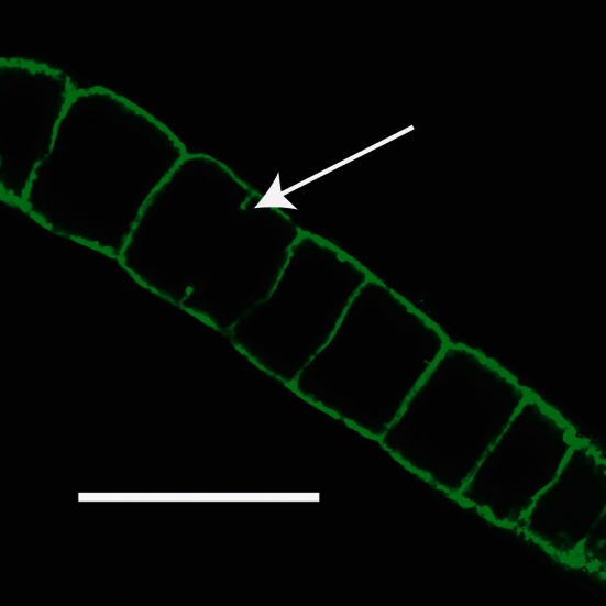
Confocal image of a Juan de Fuca filament stained with FITC showing the large internal vacuoles; Bar, 100 μm. Arrow indicates partial cross-wall in longest cell, which is presumed to be in the process of dividing
Official websites use .gov
A
.gov website belongs to an official
government organization in the United States.
Secure .gov websites use HTTPS
A lock (
) or https:// means you've safely
connected to the .gov website. Share sensitive
information only on official, secure websites.
Confocal image of a Juan de Fuca filament stained with FITC showing the large internal vacuoles; Bar, 100 μm. Arrow indicates partial cross-wall in longest cell, which is presumed to be in the process of dividing