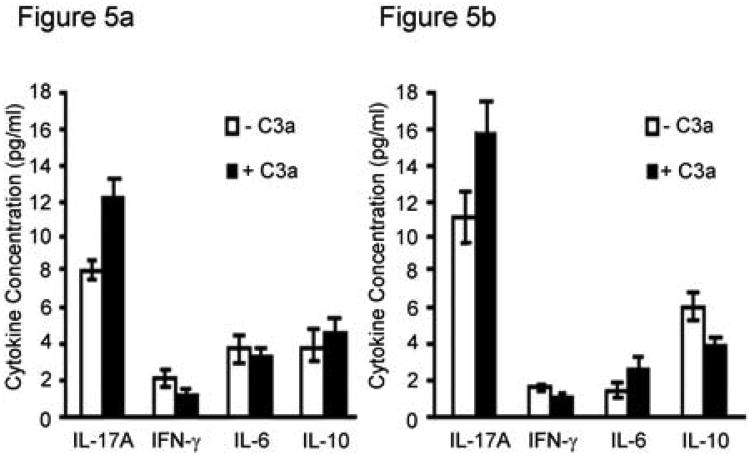
Figure 5. C3a effects on cytokine production and lymphocyte proliferation.
A. CD3+ splenic T lymphocytes (3 × 10⁵) derived from C57BL/6 that received lung allografts from C57BL/10 mice were incubated with T cell-depleted splenocytes from C57BL/10 mice as a source of antigen presenting cells (3 × 10⁵) in the presence and absence of C3a (10 ng/ml). B. Pure CD3+ T cells from col(V)-immunized mice (C57BL/6, 3 × 10⁵) were incubated with T cell-depleted splenocytes from C57BL/6 mice as a source of antigen presenting cells (3 × 10⁵) in the presence and absence of C3a (10 ng/ml). Conditioned medium was assessed for cytokines by cytometric bead array after 72 hour incubation. Levels of cytokines from wells of T lymphocytes alone or antigen presenting cells alone were below the level of detection. Values represent averages ± S.D. of three independent experiments.

