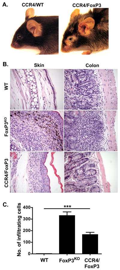
Figure 4. Characteristics of mixed CCR4/FoxP3 BM chimeras.
(A) A representative CCR4/FoxP3 chimeric mouse (right) displaying skin inflammation, erythema, alopecia and crusting on the eyes, ears and back eleven weeks after BM reconstitution. CCR4/WT chimeras (left) appeared phenotypically normal. (B) Cutaneous inflammation in CCR4/FoxP3 chimeras. Representative hematoxylin- and eosin-stained sections of skin (left column) and colon (right column) from WT (upper panel), FoxP3KO mice (middle panel), and CCR4/FoxP3 chimeras (lower panel) eleven weeks after BM reconstitution. (C) Infiltrating cells per 0.01 mm2 determined by evaluation of two hematoxylin- and eosin-stained sections per mouse (5 mice per group) eleven weeks after BM reconstituton. ***p < 0.0001

