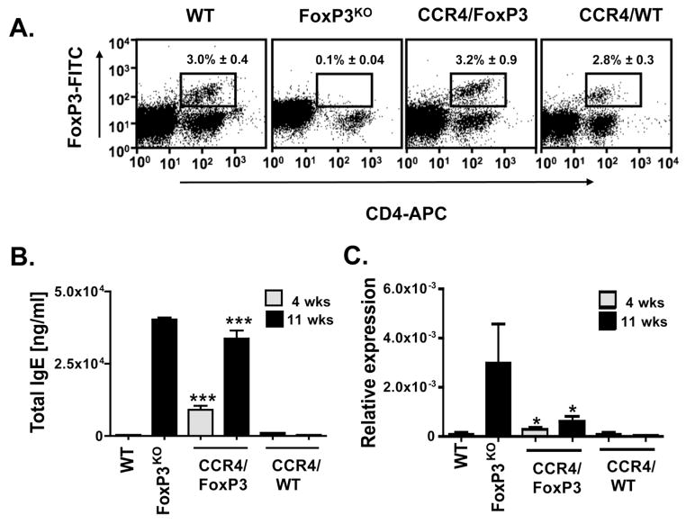
Figure 5. Characteristics of CCR4/FoxP3 chimeras.
(A) Splenocytes were stained for FoxP3, CD4 and CD25 and analyzed via flow cytometry eleven weeks after BM reconstitution. Representative dot plots are shown. Numbers indicate the average percentage of FoxP3+ and CD4+ double positive splenocytes ± SEM (n = 3). The average percentages of FoxP3+ CD4+ of total CD4+ cells constituted 19.5% ± 2.2 for WT and 32.5% ± 0.1 for CCR4/FoxP3 chimeras. (B) Serum IgE levels in WT, FoxP3KO mice and CCR4/FoxP3 and CCR4/WT chimeras four weeks (hatched column, n = 31 per group) and eleven weeks (black column, n = 3 per group) after BM reconstitution. (C) IL-4 mRNA measured by quantitative real-time PCR in skin of WT, FoxP3KO, CCR4/FoxP3 and CCR4/WT chimeras four weeks (hatched column, n = 12 per group) and eleven weeks (black column, n = 6 per group) after BM reconstitution. Relative expression of IL-4 is represented in relation to GAPDH. *p < 0.05, ***p < 0.0001

