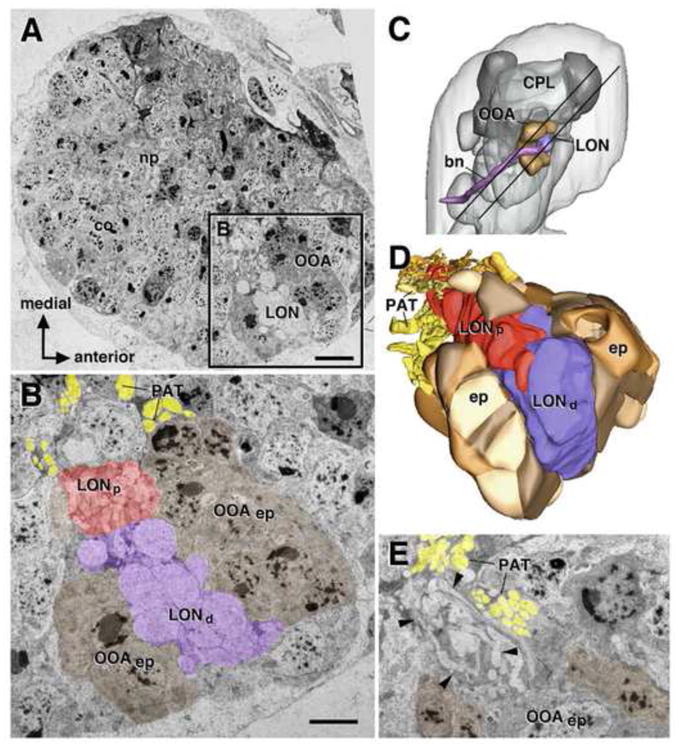
Fig. 3.

Topology of larval optic neuropil (LON) as part of series of contiguous transmission electron microscopy (TEM) images. A: Montage of TEM images of one L1 larval brain hemisphere (co cortex; np neuropil; OOA outer optic anlage). B: High magnification of section of larval visual system, boxed in (A). Shown are epithelial outer optic anlage (OOAep; rendered brown), distal LON (LONd; purple), defined by presence of large diameter photoreceptor terminals (arrows), proximal LON (LONp; red), axon tracts of primary lineages flanking optic anlage (primary axon tract; PAT; yellow). C: 3D digital model of L1 larval brain hemisphere, lateral view. Neuropil compartments (dark gray; CPL centro-posterior lateral compartment) and larval visual system (rendered in same colors as in Fig. 1A, B) are shown as landmarks. Parallel lines demarcate the brain “slice” included in the TEM stack that was used for the analysis of the larval optic neuropil in this paper. D: 3D digital model of LON and OOA, reconstructed from TEM stack. The model is shown at an orientation that is used most frequently in the following figures of this paper: the view is from dorso-posterior (indicated by red arrow in (C), exposing the LONd and LONp, enclosed by the hemicylindrical outer optic anlage. E: TEM image of proximal LON (boundaries indicated by arrowheads) at higher magnification. Scale bars: 5μm (A), 2 μm (B, E).
