
Fig. 5.
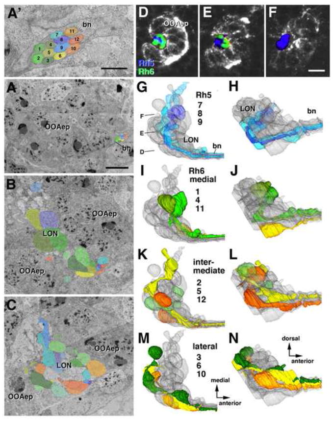
Topographically ordered projection of larval photoreceptor terminals in larval optic neuropil (LON). A–C: TEM sections of larval photoreceptor terminals and LON at different anterior-posterior levels, indicated by lines in panel H. The twelve individual photoreceptor axons are rendered in different colors and arbitrarily annotated by numbers, shown in (A′). A, A′: Bolwig’s nerve (bn) as it contacts the outer optic anlage (OOAep). Presumed Rh5-positive axons (shades of blue and purple) occupy a centro-medial position within nerve; Rh6-positive axons (shades of green to orange) are peripherally. B: Section of central part of LON. C: Section of posterior part of LON. D–F: Single sagittal confocal sections of early larval visual system (OOAep labeled by anti-DE-cadherin antibody). Planes of sections are indicated in panel G. Rh5-positive and Rh6-positive photoreceptor axons are differentially labeled by rh5-GFP and rh6-GFP. Note that Rh5-positive terminal axons (blue) occupy a posterior location in LON (arrowhead in E), and terminate mainly in proximal LON (F), which does not receive terminal axons of Rh6-positive axons (green). G–N: 3D digital models of LON (gray) and individual photoreceptor axon terminals, rendered in same colors as used in TEM images in A–C). Models of left column (G, I, K, M) present dorsal view; right column offers lateral views. G, H: three presumed Rh5-positive photoreceptor terminal axons, recognizable by their posterior position in LON (arrowhead in H) and termination in proximal (i.e., medial) LON (arrow in G). I, J: Rh6-positive receptor axons 1, 4, 11, traveling medially in Bolwig’s nerve (A′), have terminal boutons in anterior LON. K, L: Rh6-positive axons 2, 5, 12, located at intermediate levels in nerve, terminate preferentially in central domain of LON. M, N: Lateral Rh6-positive axons 3, 6, 10 terminate in posterior LON. Scale bars: 0.5μm (A′), 1 μm (A), 2 μm (B, C, D), 3 μm (D, E, F).