
Fig. 7.
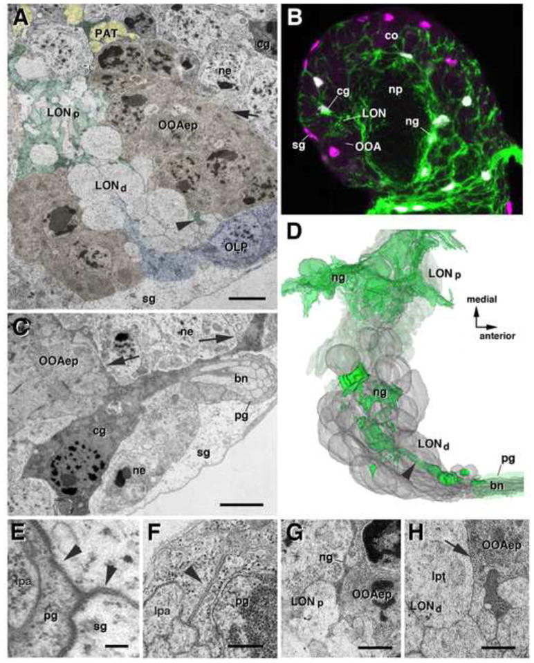
Glial element of the larval optic neuropil (LON). A: TEM section showing outer optic anlage epithelium (OOAep) and LON (LONd: distal LON; LONp: proximal LON). OOA epithelium is shaded brown; glial processes in green; optic lobe pioneers (OLP) in blue; primary axon tracts (PAT) in yellow. Arrow points at interface between apical membranes of OOA epithelial cells and neurons (ne), which lacks any glial layer. Surface glia (sg) surrounds brain surface, including OOA and optic lobe pioneers (OLP). Note cortex glial cell (cg) directly adjacent to OOA and LON, cortex neuron cell bodies (ne) (A, C). B: Single confocal section of first instar larval brain hemisphere, labeled with Nrv2-Gal4 driven GFP (green; glial cell bodies and processes) and anti-Repo (glial nuclei, magenta).. sg have repo-positive nuclei, but do not express the Nrv2 driver. Neuropil glia (ng) surrounds central brain neuropil (np), cortex (co). C: TEM section showing lateral part of the OOAep and brain cortex. Electron-dense cortex cg emits processes in between neurons of cortex (arrowheads). Note also Bolwig’s nerve (bn), surrounded by peripheral glial sheath (pg), which contacts both surface glia (sg) and cortex glia. D: 3D digital model of LON (gray) and glial processes present in LON (green). Note relative scarcity of glial lamellae in distal LON, compared to proximal LON. Arrowhead in D (and A) points at straight glial process that extends from the entry point of the bn to the center of the LON. E–H: TEM images showing details of glial structure at high magnification. E: septate junctions (arrowheads) between pg (surrounding larval photoreceptor axons, lpa) and sg. F: Auto-septate junction (arrowhead) formed by mesaxonal peripheral glia around photoreceptor axons of bn. G: Glial lamella (ng) between OOAep and neurites of LONp. H: Interface between LONd, containing boutons of larval photoreceptor terminals (lpt), and OOA epithelium. Note absence of glial sheath (arrow).