
Fig. 6.
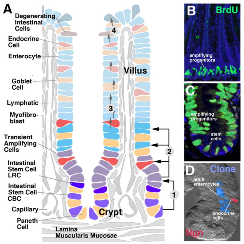
Cell replacement in the mammalian small intestine. A: Schematic depiction of intestinal villus and crypt, showing distribution of stem cells, transient amplifying cells, and differentiated cell types. Numbered arrows indicate cell movements: (1) Stem cells at crypt bottom give rise to transient amplifying cells moving towards crypt neck; (2) Transient amplifying cells produce all cell types of adult intestine; (3) Postmitotic cells stream towards tips of villi; (4) Damaged postmitotic cells at tips are sloughed off into intestinal lumen. B (low magnification) and C (high magnification): BrdU-labeled proliferating cells in crypt of mouse intestinal epithelium (from Aiken and Roth, 1992, with permission). D: Clone derived from labeled progenitor, containing enterocytes and one endocrine cell ([abeled with probe against neurogenin (Ngn; red); from Bjerknes and Cheng, 2006, with permission].