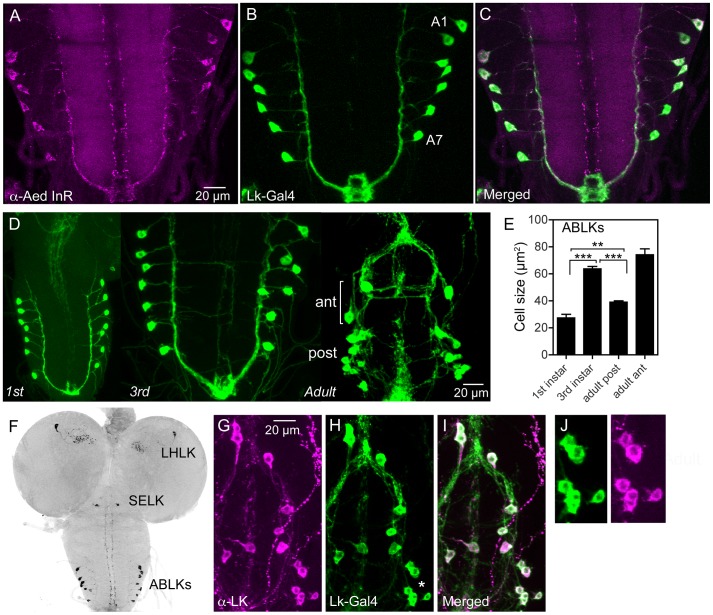
Figure 1. Distribution of insulin receptor protein in the larval CNS of Drosophila and growth of neurosecretory cells.
A–C An antiserum to part of the insulin receptor (InR) of the mosquito Aedes aegypti labels neurosecretory cells (designated ABLKs) in the abdominal ganglia of the third instar larva that also produce the neuropeptide leucokinin (LK), visualized here with Lk-Gal4 driven GFP. Seven pairs of LK neurons coexpress the markers in abdominal segments A1–A7. D The cell bodies of LK neurons (ABLKs) in A1–7 grow substantially from first to third instar larvae, but not during pupal development. Thus, the posterior neurons (post) in the 3 d old adult fly (corresponding to ABLKs) are even smaller than in the late larva. However a set of 8 anterior adult-specific LK neurons (ant) have larger cell bodies than the ABLKs. LK neurons are visualized by Lk-Gal4-driven GFP. E Quantification of cell body sizes in larvae and 3 d old adult flies. The ABLKs (14 posterior neurons) are significantly larger in 3rd instar larvae than in both 1st instar and 3 d old adults (unpaired Student's T-test; **p<0.01, ***p<0.001; n = 5–6 animals for each developmental stage). F Overview of the larval CNS (3rd instar) with LK-immunolabeled neurons, LHLK, SELK and ABLKs. G–J The immunolabeling of LK neurons with anti-LK (F) is a good marker for cell body size since it produces the same outline as membrane-targeted GFP (mcd8GFP) driven by the Lk-Gal4 (G). The posterior cell bodies marked by an asterisk are shown in higher magnification in J.

