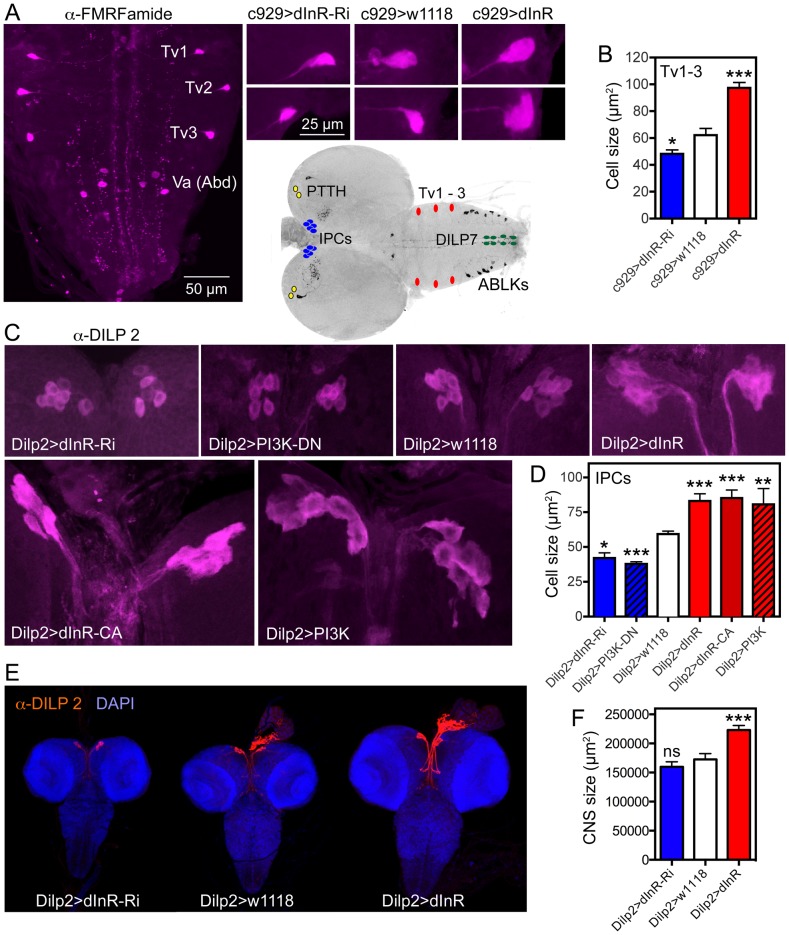
Figure 7. Dimm expressing neurons respond to manipulations of the dInR and PI3K.
The inset at the center shows the localization in the CNS of third instar larva of neurons depicted in Fig. 6, 7 and Fig. S11. A Using the c929-Gal4 to drive dInR constructs, and an antiserum to FMRFamide, we monitored the cell body size of the thoracic Tv1 – 3 neurons, known to express Dimm. As seen in the smaller panels the cell bodies are smaller after dInR-RNAi and larger after dInR over expression. B Quantification of Tv1–3 cell body sizes (*p<0.05, ***p<0.001, n = 6–8 animals for each genotype from 3 crosses; unpaired Student's T-test). C and D Also the cell bodies of the insulin producing cells (IPCs) of the larval brain respond significantly to manipulations of the dInR as well as PI3K. A dominant negative form of PI3K (PI3K-DN) was used. D Quantification of cell body sizes of IPCs (*p<0.05, **p<0.01, ***p<0.001, n = 6–16 animals for each genotype from 3 crosses; unpaired Student's T-test). E Overexpression of dInR on IPCs also affect IIS and has systemic effects on growth of the CNS. F Quantification of the dInR mediated growth of the CNS. Only overexpression of dInR produces a significant change of CNS size (***p<0.001; unpaired Student's T-test; n = 10–12 animals for each genotype, from 3 crosses).

