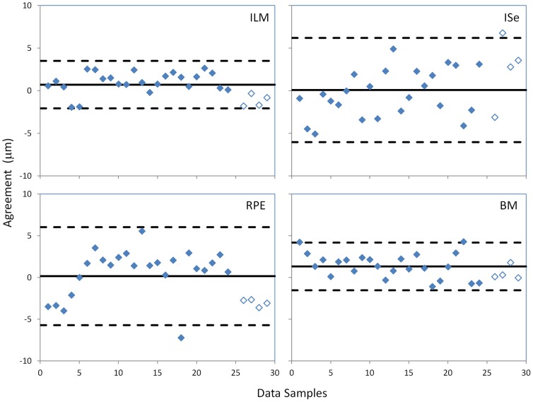
Figure 4. Agreement in segmented layer locations between the manual and EdgeSelect methods.
Solid lines indicate the mean difference of the agreement between the two methods and the dashed lines indicate mean ± 2 standard deviations. The filled symbols are data points from patients and the open symbols are from normal subjects. ILM: inner limiting membrane; ISe: the inner-segment/ellipsoid interface; RPE: the retinal/retinal pigment epithelium interface; BM: the Bruch's membrane.

