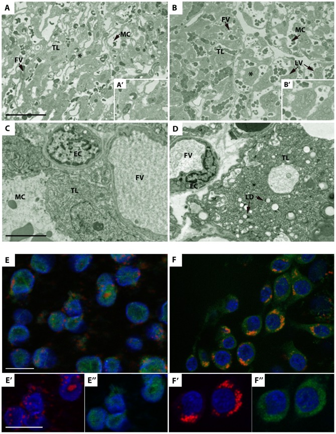
Figure 4. Morphological analysis of placentas on D28 of gestation and distribution of lipid droplets on D6 in blastocysts.
(A-B) Light microscopy morphological analysis of placentas on D28 from C and HH fetuses. Numerous light vesicles (LV) are located in the trophoblastic layer (TL) of HH (B) compared to C placenta (A). A’ and B’ represent the high magnification of the zone indicated by a star. Scale bar: 100 µm. (C-D) Transmission electron microscopy of placentas on D28 of gestation from C and HH fetuses. Light vesicles were identified as lipid droplets (LD) in the trophoblastic layer (TL) of HH (right panel) compared to C placenta (left panel). EC: Endothelial cell; FV: Fetal Vessel; MC: Maternal Compartment. Scale bar: 5 µm. (E-F) Distribution of lipid droplets on D6 in C and HH blastocysts Fluorescent immunodetection of adipophilin (green), Nile red (red), and DNA (blue) from C (E) and HH (F) blastocysts. In HH blastocysts, abnormal accumulation of lipid droplets is observed around the nuclei (F’), colocalized with adipophilin staining (F’’), in contrast to C blastocysts (E-E”). Scale bar: 20 µm.

