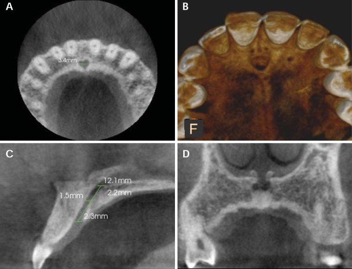
Fig. 1.
The nasopalatine canal on CBCT images. A. An axial section at the level of the incisive fossa shows the medio-lateral diameter of the incisive fossa. B. A three-dimensional reconstructed image of the nasopalatine canal. C. A sagittal section shows the measurements of the length of the nasopalatine canal and the antero-posterior diameter of the canal at the nasal fossa, mid-level, and hard palate. D. A coronal section shows two openings of Stenson's foramina.

