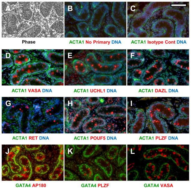
Figure 1. Localization of SPG antigens in pre-pubertal canine testis.
(A) Phase image of tissue after deparaffinization. (B–L) Duel label probes in which putative SPG antigens (red) and somatic cell antigens (green) were marked. Putative SPG antigens (red) were probed with either rabbit (C–I) or goat (J–L) primary antibodies as indicated, followed by the appropriate Alexa-594 conjugated donkey secondary antibody. Somatic cells (green) were labeled either with mixed mouse antibodies against αActin and vimentin, (B–I) or GATA4 (J–L) followed by an anti-mouse Alexa 488 conjugate. The specific antibodies and dilutions used are shown in Supplemental Table I. Controls for background labeling were done identically except that they included either no primary antibody (B) or a rabbit isotype control (C). Specific antibodies and dilutions are listed in Supplemental Table I. Nuclei were counterstained blue with Hoechst 33342 in some sections (B–I). Bar = 100 um.

