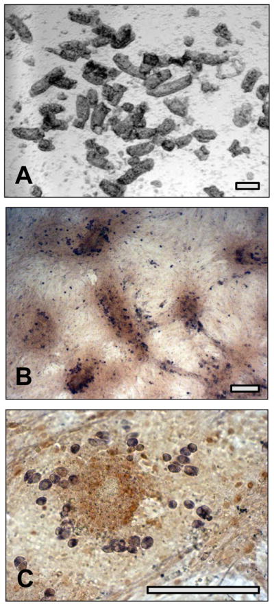
Figure 3. Emergence of SPG as a “loose cell” population in cultures of total testis cells.

(A) Bright field image of partially digested seminiferous tubules used to initiate culture. (B) Immunohistochemistry for VASA (purple) and GATA4 after 3 days culture of total testis cells. Note remnants of tubule pieces (dark brown) are firmly attached to substratum. (C) Higher power image of culture shown in (B). The VASA-positive cells are rounded and loosely scattered on top of the dense fibroblast-like lawn. Bars = 100 um.
