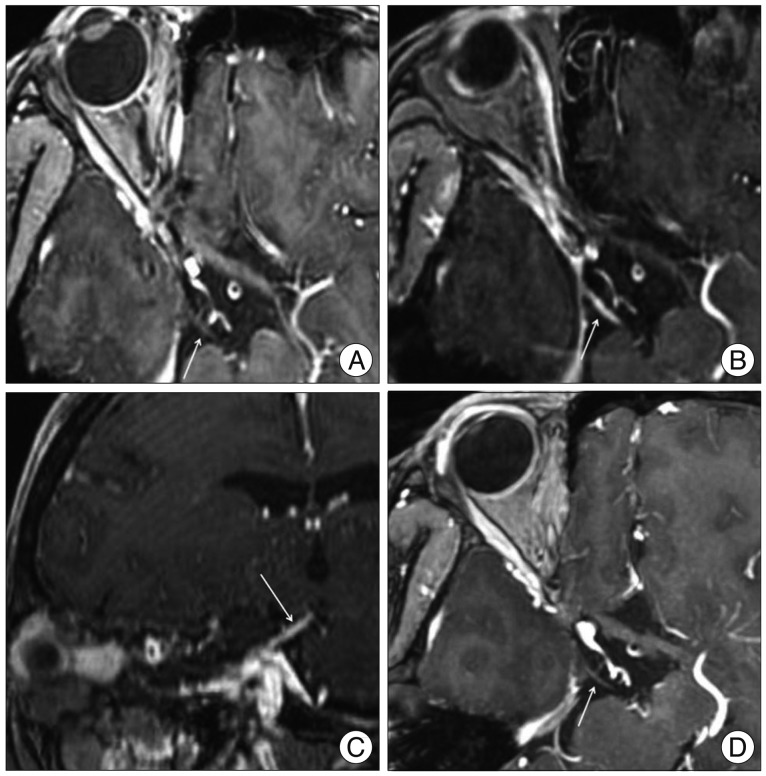
Fig. 3.
Multiplanar reconstruction magnetic resonance images using contrast-enhanced spoiled gradient-recalled sequences clearly depict the oculomotor nerves (ONs) (arrows) in head trauma patient. A : Initial image shows focal swelling of the right ON at the posterior petroclinoid ligament, but no gadolinium enhancement. B and C : Axial and sagittal reconstruction images obtained 16 days after the injury reveal diffuse thickening and enhancement of the cisternal portion of the right ON. D : Follow-up study confirms virtually complete resolution of the enhancement of the nerve itself.

