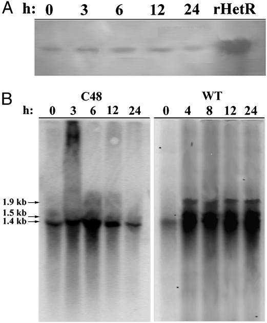
Fig. 6.
Analyses of the hetRc48a expression in C48 after shifting from a nitrogen replete condition to a nitrogen-deprived condition. (A) Immunoblotting analysis of the HetR proteins. Total cellular protein extracts were prepared from cells at the times indicated after nitrogen step-down and separated by SDS/PAGE before electrophoretic transfer to a polyvinylidene difluoride membrane. HetR was detected as described in Fig. 2. (B) Northern blotting analysis of the expression of hetR and hetRc48a. Total RNA was isolated from the cells at the times indicated after nitrogen deprivation and separated with 1.5% agarose gel electrophoresis before transfer to a nylon membrane. The mRNA of hetR and hetRc48a was detected by using a 32P-labeled hetR probe. The transcript sizes are shown on the left. The smear on top of the 3-h lane in C48 could probably be resulted from unremoved polysaccharides in the RNA sample.

