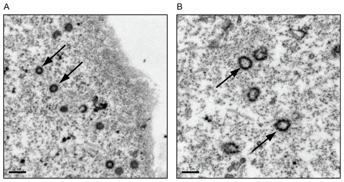
Figure 2. Verification of viral particles shapes by transmission electron microscopy.
COS-1 cells were transfected with pSARM-X wild type provirus (A) or pSARM-GagGFP-M100A (B). 24 hours after transfection were cells fixed in a 2.5% glutaraldehyde fixative for 2 hours at 4 °C. The cells were washed and subsequently fixed in 1% osmium fixative. The cells were then dehydrated with ethanol, followed by infiltration with 100% ethanol and Epon resin for at least one hour. Cells were then embedded in fresh Epon resin at 60°C for 48 hours then cut into ultrathin sections and picked up on a copper grid. The arrows in (A) point to WT intracellular capsids of homogenous shape, with a diameter of approximately 100 nm. The arrows in (B) point to GagGFP-containing intracellular capsids having a beads-on-a-string shape and varying diameters greater than 100 nm. Bar = 200 nm.

