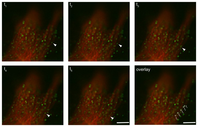
Figure 6. Real-time imaging of GFP labeled virus-like particles along the microtubule.
M-PMV expressing CMMT cells were cotransfected with pSARM-GagGFP-M100A and mCherry-tubulin and imaged 24 hours later in real-time. Images were captured at consecutive intervals of 1 second for one minute in one focal plane. Scale bar= 1 µm. Real time imaging of these frames is shown in movie S2.

