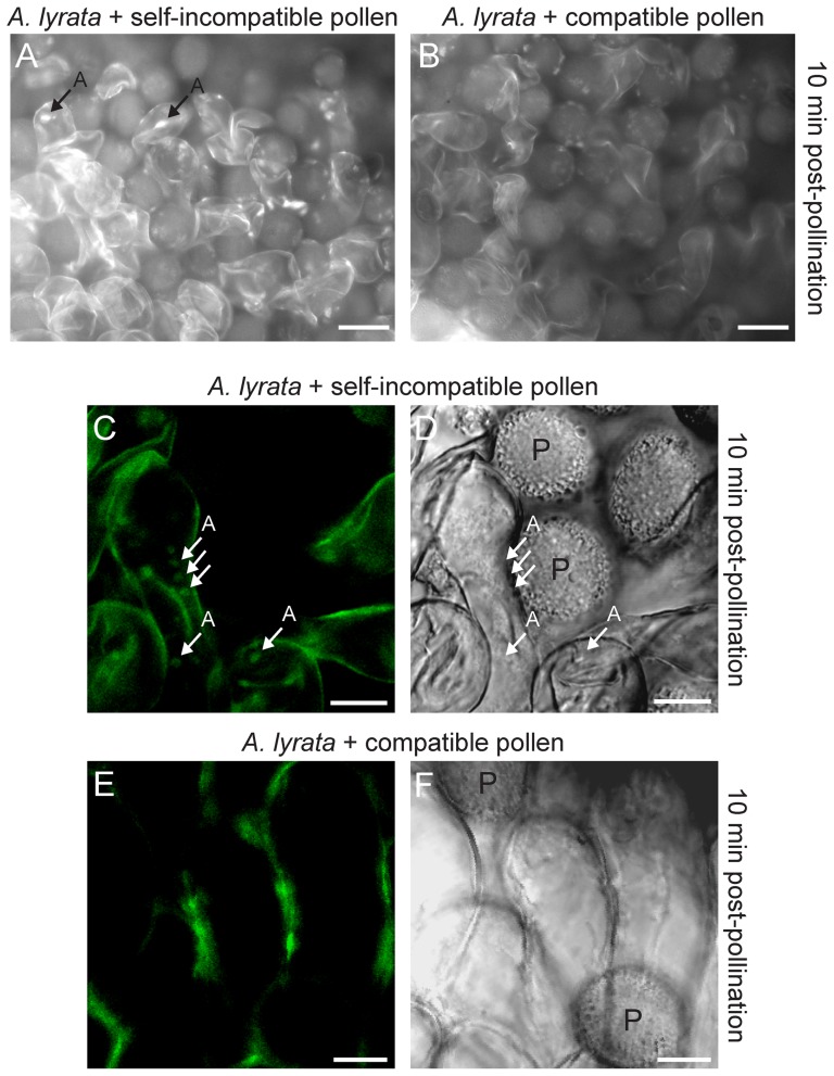
Figure 8. Autophagosomes in A. lyrata stigmatic papillae in response to self-incompatible pollen.
(A, B) Florescence microscopy images of MDC stained A. lyrata stigmatic papillae at 10 min post-pollination. Fluorescent signals that may represent autophagosomes were seen in the A. lyrata stigmatic papillae following a self-incompatible pollination (A) in 10/10 samples, but not observed after a cross-compatible pollination (B) in 10/10 samples. (C-F) Confocal microscopy images of transgenic A. lyrata GFP:ATG8a stigmatic papillae at 10 min post-pollination. GFP:ATG8a is a marker for autophagy induction, and GFP signals marking potential autophagosomes were observed in the stigmatic papillae following a self-incompatible pollination (C) in 10/10 samples (corresponding DIC image is shown in D). Punctate GFP signals were not detected within the stigmatic papillae following a cross-compatible pollination (E) in 10/10 samples (corresponding DIC image is shown in F). All samples, including wild-type untransformed A. lyrata stigmatic papillae showed background fluorescence from the cell wall. A = autophagosomes; P = pollen. Scale bars (A, B) 50 µm; (C-F) 10 µm.

