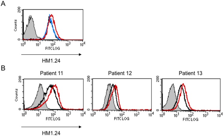
Figure 3. Len enhanced the HM1.24 expression on primary MM cells in the presence of effector cells.
(A) RPMI 8226 cells were cultured for 48 hours in the absence (black) or presence of 3 µM Len alone (blue), or 3 µM Len plus PBMCs from a healthy donor using membrane filters to avoid cell contact (red). (B) BMMCs from MM patients (no. 11, 12, and 13) were cultured for 48 hours in the absence (black) or presence of 3 µM Len (red). Thereafter, the MM cells were stained with control FITC-labeled mouse IgG or FITC-labeled HM1.24 mAb, and PE-labeled CD38 mAb, and analyzed by flow cytometry. MM cells were gated according to a side scatter (SS) and CD38 expression, and analyzed for their HM1.24 expression.

