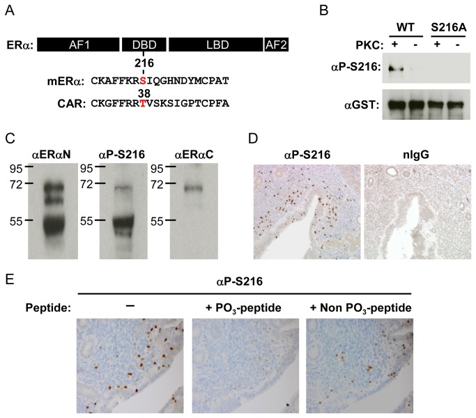
Figure 1. Phosphorylation of ERα at serine 216.
(A) Map to show localization of serine 216 in ERα and threonine 38 in CAR. (B) In vitro phosphorylation of serine 216. Purified glutathione S transferase (GST)-mERα wild type (WT) and GST-S216A were incubated with or without protein kinase C (PKC) and Western blots were performed using an anti-P-Ser-216 (αP-S216) or anti-GST antibody. (C) Western blot analysis of whole extracts (20 μg protein/well) prepared from the mouse uterus at the estrus stage with αP-S216 or an anti-ERα antibody (αERαN or αERαC). (D) Uterine sections were immune-stained using αP-S216 antibody or normal IgG. (E) Uterine sections to determine staining specificity of αP-S216. αP-S216 antibody was mixed with phospho- or non-phospho-peptides prior to incubation with sections.

