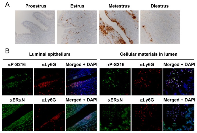
Figure 3. Phosphorylated ERα-expressing neutrophils during the estrous cycles.
(A) Mouse uterine sections (at the proestrus, estrus, metestrus or diestrus stage) were stained by an anti-Ly6G antibody (αLy6G). (B) Sections were prepared from the mouse uterus at the metestrus stage and subjected to fluorescence double staining with αLy6G and either αP-S216 or αERαN. Immunoreactions were visualized as described in the legend of Figure 2. DAPI stains nuclei.

