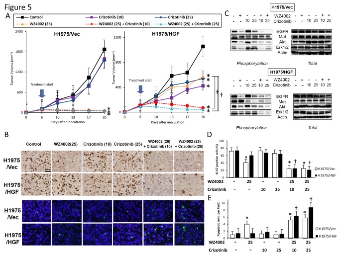
Figure 5. Crizotinib combined with mutant-selective EGFR-TKI overcomes multiple resistances to EGFR-TKI in vivo.
(A) SCID mice-bearing H1975/Vec- or H1975/HGF- tumors were administered WZ4002 (25 mg/kg) and/or crizotinib (10, 25mg/kg) once daily for 6 to 20 days. Tumor volume was measured using calipers on the indicated days. Mean ± SE tumor volumes are shown for groups of 5 mice. *, P < 0.05 versus control; ✝, P < 0.05 versus WZ4002 by one-way ANOVA. (B) H1975/Vec- or H1975/HGF- tumors were resected from the mice 3 hours after administration of WZ4002 (25mg/kg) and/or crizotinib (10, 25 mg/kg), and the relative levels of proteins in the tumor lysates were determined by western blot analysis. (C) Representative images of H1975/Vec- and H1975/HGF- tumors immunohistochemically stained with antibodies to human Ki-67, and stained with both DAPI (nuclear stain) and TUNEL (FITC). Bar, 200 μm. (D) Quantification of proliferative cells, as determined by the Ki-67-positive proliferation index (percentage of Ki-67-positive cells). Quantification of apoptotic cells, as determined by the TUNEL assay as described in Materials and Methods. Columns, mean of five areas; bars, SD *, P < 0.05 versus of H1975/Vec-tumors; ✝, P < 0.05 versus H1975/HGF-tumors by one-way ANOVA.

