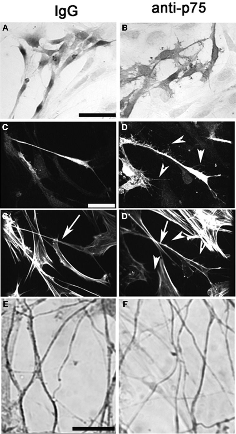
Figure 7.
Perturbation of p75NGFR signaling in vitro alters morphology of OECs and causes defasciculation of olfactory fibers. Nasal explants at 6div following treatment with p75NGFR blocking antibody (1:6000, B,D,F) and with rabbit IgG mock, 1:6000, (A,C), Explants were immunocytochemically-stained for S-100 (A,B), S-100 and Phalloidin (C,D) or peripherin (E,F). (A–D) Disruption of p75NGFR signaling alters morphology of OECs The morphology of S100-positive cells located distal to the explant's tissue mass were larger, flatter, and exhibit more processes and neurites (B,D) as compared to control group (A,C). Staining for actin (phalloidin) revealed that these processes had sparse actin filaments (D′, arrowheads). OECs in the treated group also appeared to have denser cortical actin along the axis of the cell body (C′ vs. D′, arrow). Finally, disruption of p75NGFR increased olfactory fiber branching and decreased the optical density of peripherin-positive fibers, suggesting defasciculation had occurred. Scale bars: = 250 μm.

