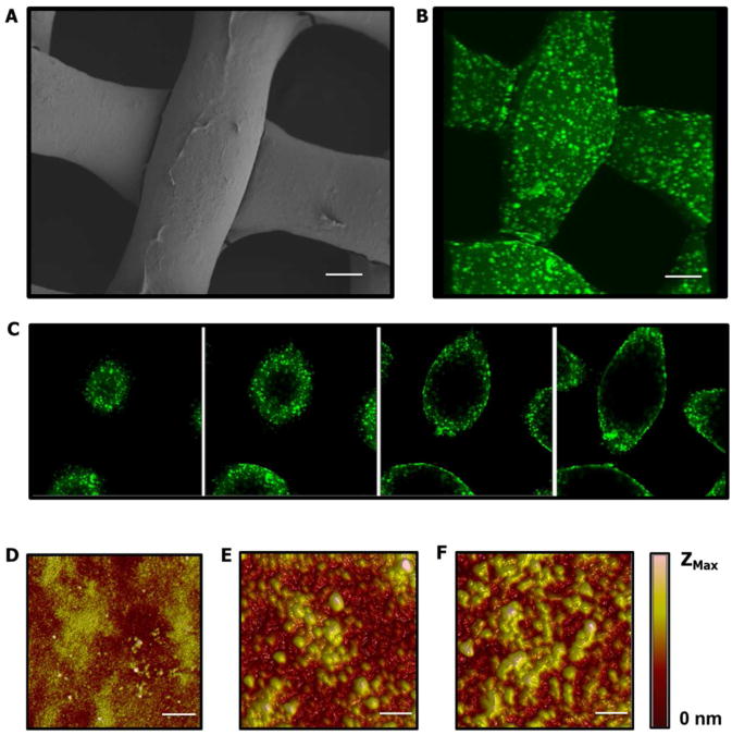
Figure 4.
Characterization of Laponite® containing LbL film coating on Tegaderm® substrate. (A) SEM imaging of film coated substrate, scale bar = 25 μm. (B) Three dimensional projection of fluorescent confocal imaging of film coated substrate using AlexaFluor 488-labeled siRNA. Scale bar = 25 μm. (C) Selected confocal images used to generate projected image. Images were selected at 8 μm steps to show the conformal nature of the film coating. (D-F) Atomic force micrographs at 5, 15, and 25 architecture repeats respectively, Zmax = 57nm (D), 138nm (E), and 182nm (F), scale bar = 5 μm.

