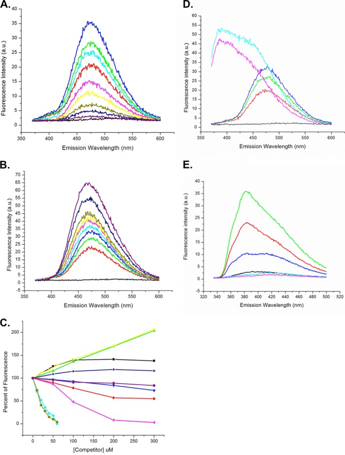
FIGURE 6.
Ligand binding by Ara h 8. A, titration of 40 μm ANS (black) with Ara h 8: 20 μm (red), 30 μm (green), and 40 μm (blue). Also shown is titration of 40 μm ANS, 40 μm Ara h 8 with quercetin: 10 μm (cyan), 20 μm (pink), 30 μm (yellow), 40 μm (olive), 50 μm (navy), and 100 μm (purple). B, titration of 40 μm ANS (black) with Ara h 8: 20 μm (red), 30 μm (green), and 40 μm (blue). Titration of 40 μm ANS, 40 μm Ara h 8 with daidzein: 20 μm (cyan), 40 μm (pink), 60 μm (yellow), 100 μm (olive), 200 μm (navy), and 300 μm (purple). C, compilation of fluorescence data. 100% was the value of fluorescence for 40 μm ANS, 40 μm Ara h 8. The values for increases or decreases in fluorescence for each concentration of ligand are shown relative to the 100% value. Shown are progesterone (black), myristic acid (red), arachidic acid (green), caffeic acid (blue), quercetin (cyan), genistein (pink), daidzein (yellow), apigenin (olive), stigmasterol (navy), and zeatin (purple). D, titration of 40 μm ANS (black) with Ara h 8: 20 μm (orange), 30 μm (green), and 40 μm (blue). Shown is titration of 40 μm ANS, 40 μm Ara h 8 with resveratrol 50 μm (pink) and 100 μm (cyan). E, fluorescence of resveratrol. Shown is titration of 50 μm resveratrol (cyan) with Ara h 8: 10 μm (red) and 20 μm (green). Also shown is titration of 50 μm resveratrol, 20 μm Ara h 8 with 10 μm quercetin (dark blue) and 20 μm quercetin (light blue).

