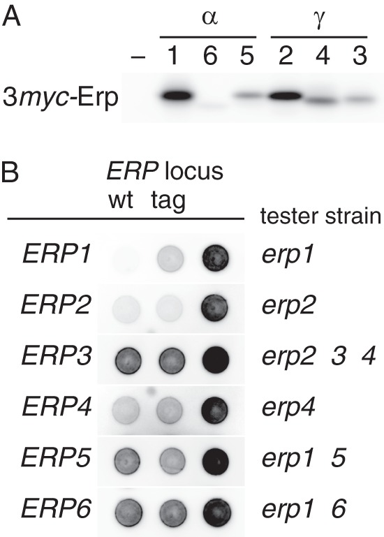
FIGURE 2.

Epitope tagging of p24α and p24γ proteins. A, detection of 3myc-tagged p24α (Erp1, -5, and -6) and p24γ (Erp2, -3, and -4). Whole-cell extracts (∼2 × 10⁶ cells) of wild-type (–) and 3myc-tagged p24 recombinant strains (1–6, the numbers correspond to the ERP gene numbers) were analyzed as in Fig. 1C. B, complementation of p24α/γ mutations by the 3myc-tagged versions of the respective genes. Tester strains (also as negative controls; Δ, right spots) used are indicated at the right side of each row. Wild-type (positive controls; wt, left spots) or 3myc-tagged (tag, center spots) ERP genes of interest were introduced to the tester strains on an integration vector. Kar2 secretion was assayed as in Fig. 1A. Blots for Erp1, Erp2, and Erp4 were overexposed for maximum clarity.
