FIGURE 6.
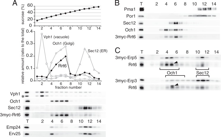
Cellular localization of Rrt6. A, sucrose density gradient fractionation of Rrt6. Cells expressing 3myc-Rrt6 were converted to spheroplasts and lysed, and cleared lysate was layered on a 25–60% (w/v) sucrose density gradient. After centrifugation at 200,000 × g for 2.5 h, fractions were collected from the top and assayed for the amounts of 3myc-Rrt6 and organelle markers. The asterisk indicates a major degradation product of Vph1. B, sucrose density gradient in the presence of 1 mm EDTA. Fractionation was done as in A, except that the lysis buffer and the gradient contained 1 mm EDTA. C, sucrose density gradient fractionation of Erp3 and Erp5. Membranes of cells expressing 3myc-Erp3 or 3myc-Erp5 were fractionated as in A. Closed triangles indicate an unrelated cross-reacting protein. Brackets show the distribution of the Golgi marker Och1 and the ER marker Sec12.

