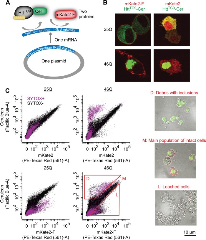
FIGURE 1.
Biosensor platform to track Httex1 aggregation inside the cells and effect of aggregation on cell viability. A, schematic of the IRES vector system employed. The vector expresses Httex1TC9, which is a biosensor of the Httex1 aggregation state that we previously showed can bind to the biarsenical dyes FlAsH or ReAsH when Httex1 is monomeric but not when it aggregates (17). Httex1TC9 is fused to Cerulean as a marker to track Httex1 expression levels. Downstream the IRES sequence is the mKate2 fluorescent protein. We created two IRES versions, one with a farnesylation targeting sequence appended to the C terminus of mKate2 (mKate2-F) and another lacking this sequence (mKate2). B, expression and imaging of the IRES constructs in Neuro2a cells with derivatives of Httex1TC9 containing different polyQ lengths. C, flow cytograms of Neuro2a cells transfected with the IRES vectors. Cells were pre-gated as indicated in Fig. 2. The gates L, M, and D were used for sorting, recovery, and visual inspection by microscopic imaging.

