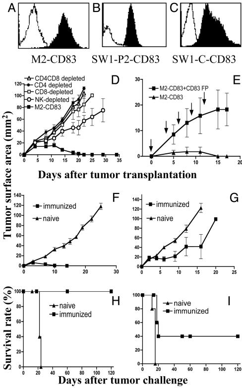
Fig. 1.
(A-C) Flow cytometry data showing expression of CD83 on transfected (filled area) versus WT (open area) cells. (D) Growth of M2-CD83 cells in mice depleted of the indicated cell populations. (E) Outgrowth of M2-CD83 cells in mice injected i.p. with 100 μg of mCD83 fusion protein (CD83 FP) at the time of tumor transplantation and repeated as indicated. The control group was injected with PBS. (F and H) Regression of M2-WT cells. (G and I) Regression of Ag104 cells in some mice immunized by live M2-CD83 cells (n = 5 mice per group).

