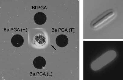
Fig. 2.
(Left) Double immunodiffusion showing reactivity of mAb F26G3 with γDPGA from B. licheniformis (Bl PGA), total γDPGA isolated from B. anthracis [Ba PGA (T)], the low molecular weight fraction of γDPGA isolated from B. anthracis [Ba PGA (L)], and the high molecular weight fraction of γDPGA isolated from B. anthracis [Ba PGA (H)]. The arrow identifies a weak precipitin line produced by a high molecular weight component of the total γDPGA. (Right) Binding of Alexa 488 mAb F26G3 (50 μg/ml) to B. anthracis when viewed by differential interference contrast microscopy to show quellung reaction (Upper) or by confocal microscopy (Lower).

