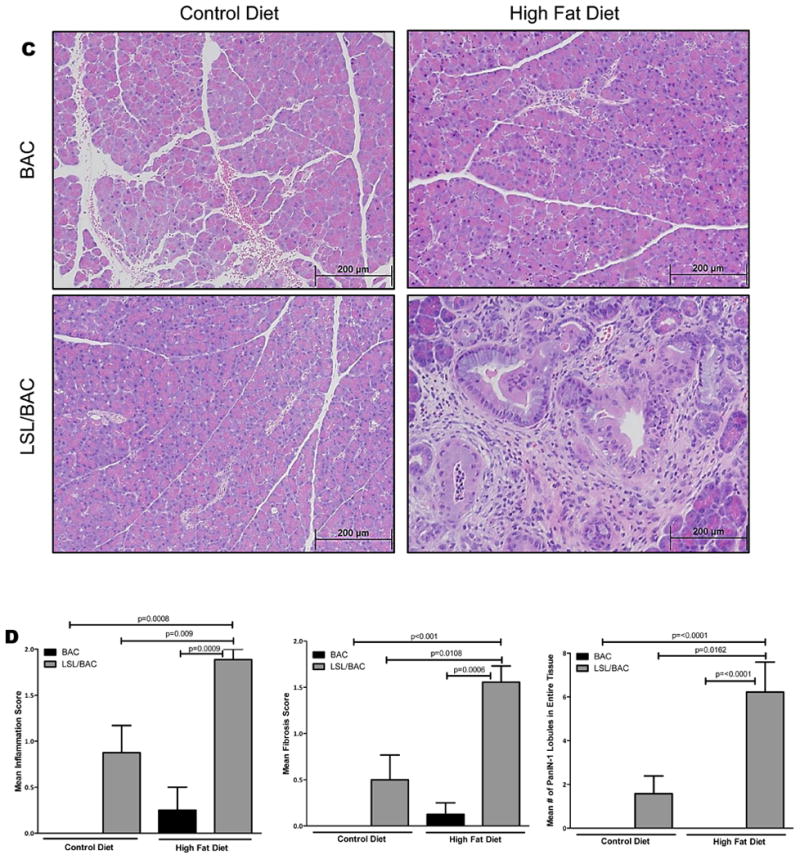
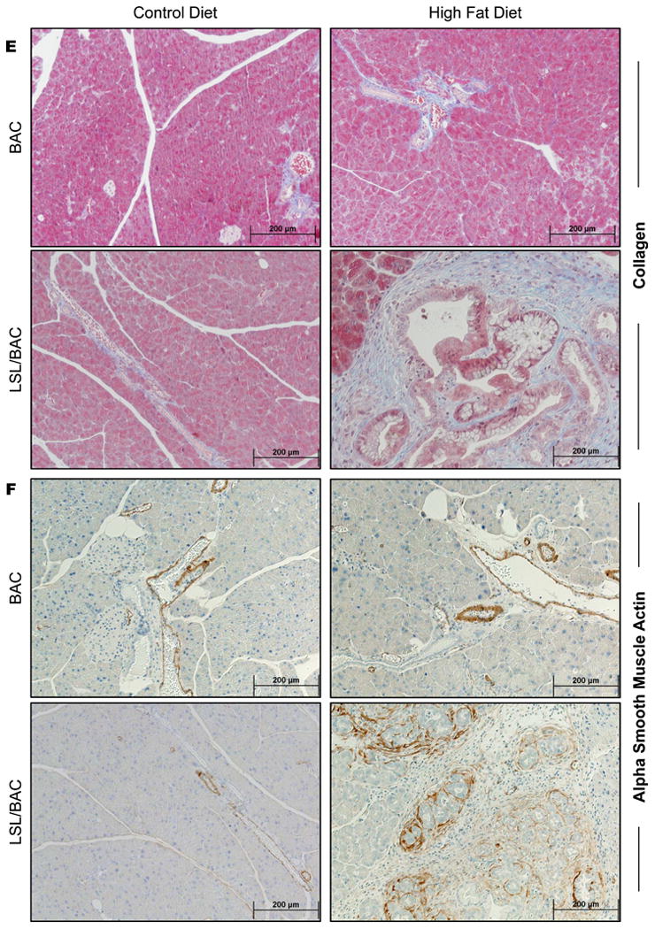
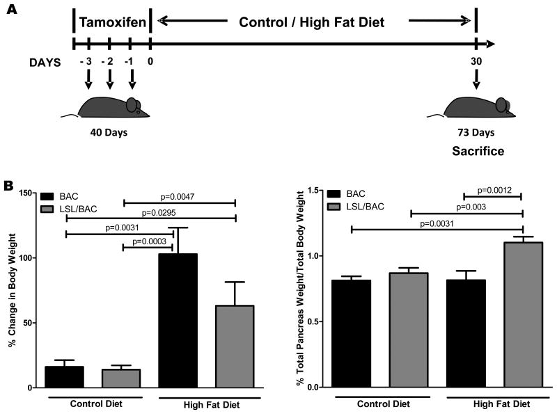
Figure 1. HFD Increased BW and PW and the initiation of the early stages of PDAC development.


(A) At 40 days of age BAC and LSL/BAC mice were induced with tamoxifen for 3 consecutive days for Cre activation and placed on CD (BAC; n=5 and LSL/BAC; n=8) or HFD (BAC; n=9 and LSL/BAC; n=10) for thirty days. (B) BW of each mouse was measured weekly and the percent change from week 1 to week 5 of treatment was calculated. In addition, PW of each mouse was measured and the percent PW/BW was graphed for each group. (C) Representative H&E stains of BAC and LSL/BAC mice on CD and HFD are shown. (D) Each tissue sample was scored according to its level of inflammation, fibrosis, and number of PanIN-1 lesions seen in the entire tissue. (E) Collagen staining was performed for BAC and LSL/BAC animals on the CD and the HFD. (F) IHC for α--SMA expression was also performed for all groups.

