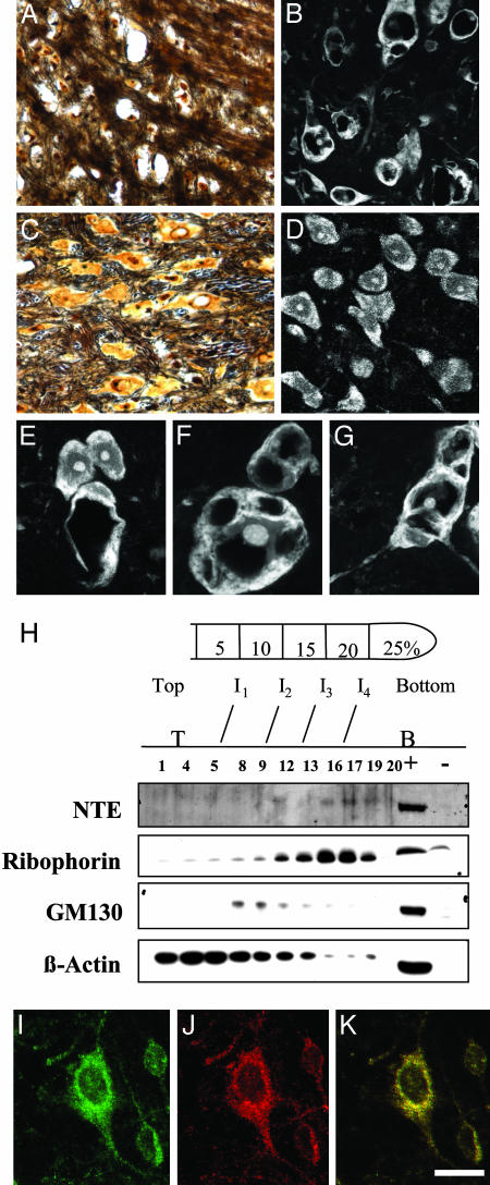
Fig. 3.
NTE localizes to the ER and its deletion results in redistribution of Nissl substance in the thalamic neurons. (A and B) Silver staining showing neuronal loss in the thalamus of a Nes-cre:NTEfl/fl mice mouse (3 months) (A) when compared to age-matched NTEfl/fl mouse (C). (E, F, and G) Fluorescent Nissl stain showing vacuolated neurons in Nes-cre:NTEfl/fl mice. (D) Normal distribution of Nissl substance in the cell body of NTEfl/fl mice. (E) Six-week-old Nes-cre:NTEfl/fl mouse at the onset of neuronal pathology showing two normal and one vacuolated neuron. (H) Subcellular fractionation of hippocampal neurons shows the presence of NTE in the ribophorin, ER-containing fraction, and not in the GM130, Golgi-containing fraction. +, positive control, cerebrum lysate; -, negative control, heart lysate; I, interface between gradients; T, top of gradient; B, bottom of gradient. Double immunofluorescence for calnexin (J) and NTE (I) in primary hippocampal neurons shows association of NTE with the ER (K). (Scale bar, 36 μmin A-C;25 μmin B-D; and 13 μmin E-G.)

