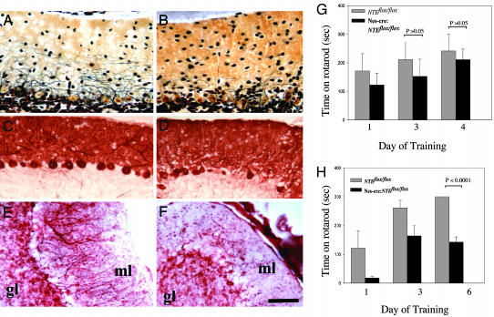
Fig. 5.
Loss of Purkinje cells and behavioral deficits caused by brain deletion of NTE. (A) Normal Purkinje cell appearance and Purkinje dendritic tree extension revealed by silver staining in the molecular layer of the cerebellum of NTEfl/fl mice. (B) Loss of Purkinje trees and reduced dendritic tree extension and complexity by silver stain of the cerebellum of Nes-cre:NTEfl/fl mice. (C) Normal number of calbindin positive Purkinje cells in NTEfl/fl mice. (D) Loss of calbindin positive Purkinje cells from Nes-cre:NTEfl/fl.(E) Normal appearance of MAP-2 positive Purkinje cell dendrites and neurons in the molecular layer of NTEfl/fl mice. (F) Loss of MAP-2 positive dendritic trees in the cerebellum. (G) No behavioral differences observed at a rotarod behavioral test measuring motor coordination between 4-week-old NTEfl/fl (n = 4) and Nes-cre:NTEfl/fl mice (n = 5), P > 0.05. (H) Evident coordination defects of 5-month-old Nes-cre:NTEfl/fl mice (n = 4), when compared to NTEfl/fl littermate controls (n = 7), P < 0.0001. GL, granular layer. (Scale bar, 50 μm.)

