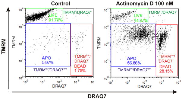
Figure 2.

Discrimination of viable, apoptotic and late apoptotic/necrotic cells based on Δψm marker tetramethylrhodamine methyl ester (TMRM) and plasma membrane permeability marker DRAQ7. Analysis was based on real-time labeling of THP-1 cells with 3 μM of DRAQ7 and 150 nm of TMRM. Bivariate dot plots DRAQ7 vs. TMRM enable discrimination of live (TMRM+/DRAQ7−; green gate), early apoptotic (TMRMlow/DRAQ7dim; blue gate) and late apoptotic/necrotic (TMRMlow/DRAQ7+; red gate) subpopulations. DRAQ7 probe was excited using 633 nm laser and logarithmically amplified fluorescence signals were collected using 660 nm long-pass filter. TMRM was excited using 488 nm laser and logarithmically amplified fluorescence signals were collected using 575 nm long-pass filter. Debris signals were excluded electronically by setting the proper low threshold.
