Figure 2.
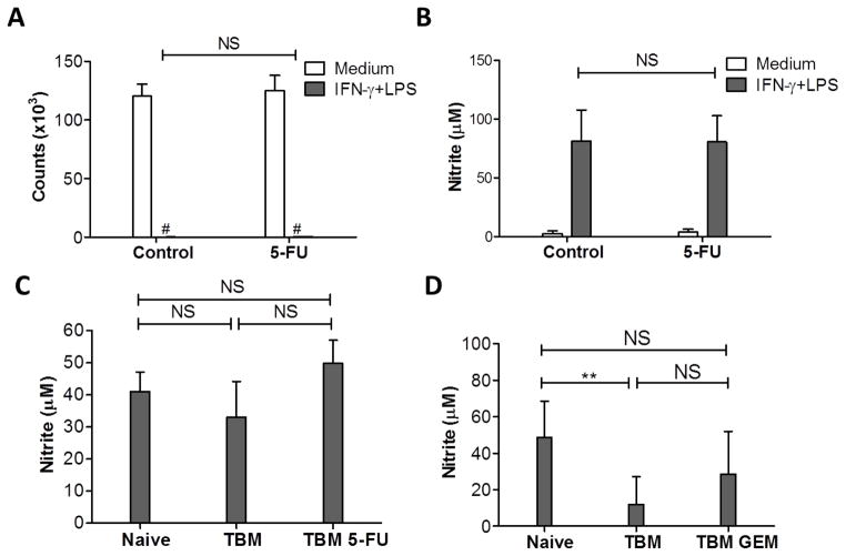
Effect of GEM or 5-FU on Mφ function. A, B: Two groups of naïve C57BL/6 mice (n=2 per group) were injected with either 50 mg/kg of 5-FU (5-FU) or DMSO (Control) i.p., and PEC were collected 5 days later. Total PEC (2x10⁵/well) and B16 tumor cells (104/well) were placed in 96-well plates with medium or stimulated with IFN-γ (10 U/ml) and LPS (1 ng/ml). C: C57BL/6 mice were injected with B16 cells i.p. on day 0, and injected with either 50 mg/kg of 5-FU (TBM 5-FU) or DMSO (TBM, control) i.p. on days 5 and 10. PECs from naïve, TBM and TBM 5-FU (n=3–4 per group) were collected on day 14. Total PEC were stimulated with IFN-γ (10 U/ml) and LPS (1 ng/ml). The results of one out of two similar experiments are shown. D: B16 i.p. TBM were injected with 120 mg/kg of GEM (TBM GEM) or PBS (TBM) i.p. on day 11. Total PEC were collected on day 14 and placed in 96-well plates with medium or IFN-γ (10 U/ml) and LPS (1 ng/ml). All plates were incubated for 48 hours. D shows a combined graph of three similar experiments (8–9 mice per group). Counts of B16 cells were measured based on thymidine incorporation (A), and NO activity was determined by nitrite level in the supernatants (B, C, D). The data are shown as Mean ± SD. # Counts <150. NS: Non-significant.

