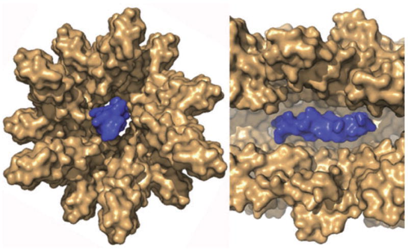
Fig. 2.

Cross sections of the proposed structure of the T3SS needle complex (brown), based on the crystal structure of MxiH modelled into a three-dimensional EM reconstruction of the needle with an helix (blue) modelled at its centre to show that the dimensions of the central channel cannot accommodate more than simple secondary structures in proteins (needle subunits, translocator proteins, effectors) that move through it. (Figure generously supplied by Janet Deane).
