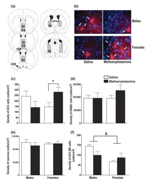
Fig. 1.
MA exposure during hippocampal development increased the density of ACh cells in the basal forebrain of adolescent female mice. (a) Representative images of the area defined as the MS/VDB/HDB and NB. (b) Representative images of ACh cells, indicated by the white arrows (red; ChAT-positive), in the MS/VDB/HDB in MA- and saline-exposed male and female adolescent mice. The blue is a DAPI stain for cell bodies. (c) MA exposure increased the density of ACh cells (ChAT-positive) in the MS/VDB/HDB in female mice compared saline-exposed female mice. This effect was not observed in adolescent male mice. (d) MA exposure did not alter the density of GABAergic cells (PVA-positive) or (e) the density of total neurons (NeuN-positive) in the MS/VDB/HDB. (f) MA exposure did not alter the density of ACh cells in the NB. Male mice had a higher density of ACh cells in the NB compared with female mice, regardless of treatment. Data expressed as mean ± SEM. n = 5–6 mice per treatment per sex. *p < 0.05 female MA-exposed versus female saline-exposed mice. &p < 0.05 male versus female mice. MS, medial septum; VDB, vertical limb nucleus of the diagonal band; HDB, horizontal limb nucleus of the diagonal band; NB, nucleus basalis; ACh, acetylcholine.

