
Figure 4. Flexible, motile extensions and cellular bridges from the non-neural ectoderm.
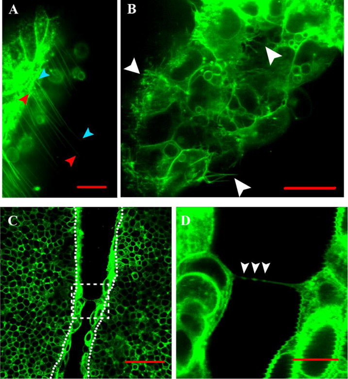
Images of the neural folds in the midbrain region. (A) Numerous long (over 50um) thin cell extensions from the non-neural ectoderm of one fold that extend across the gap towards the converging fold (not visible in this view). Scale bar 20μm.
(B) Still image from Movie 4 of the non-neural ectoderm on one fold in the midbrain region that highlights a few of the long flexible cell extensions and short bulbous flexible extensions. Scale bar 20μm.
(C) Cellular bridges connect the two juxtaposed folds when they are 20μm apart. In the boxed area, two cellular bridges connect opposing non-neural ectoderm cells. Dotted lines separate the non-neural from neural ectoderm. Scale bar 50μm.
(D) Magnification of one of the cell bridges from Figure 4C. Three structures ∼0.5 μm in diameter that are highlighted with the myristoylated Venus fluorescence reporter are present within the cellular bridge (white arrowheads). Scale bar 10μm.