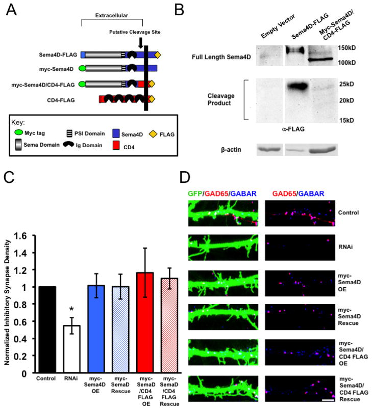
Figure 4. Sema4D promotes GABAergic synaptogenesis as a membrane bound molecule.
(a) Diagram illustrating the pertinent epitope-tagged Sema4D and CD4 chimeric constructs used in these experiments. The myc-Sema4D/CD4-FLAG fusion was constructed such that the CD4 peptide sequence replaces the putative cleavage site between the Ig domain and transmembrane domain of Sema4D (Elhabazi et al 2001).
(b) Western Blot analysis of lysates from rat hippocampal neurons infected with lentivirus containing either an empty vector, Sema4D-Flag, or myc-Sema4D/CD4-FLAG. Western blots were probed with an anti-FLAG antibody, which targets the FLAG epitope on the C-terminus of Sema4D-FLAG and myc-Sema4D/CD4-FLAG. While a 25kD C-terminal cleavage product is detectable in neurons expressing Sema4D-FLAG no cleavage product of any size ranging from 15kD to 25kD was detected in neurons expressing myc-Sema4D/CD4-FLAG.
(c) Quantification of inhibitory GABAergic synapse density at DIV14 as defined by overlapping GAD65/GABARγ2 puncta onto rat hippocampal neurons transfected with either an empty vector (“Control,” n= 18 neurons), an shRNA targeting Sema4D (“RNAi,” n= 29 neurons), overexpression of a myc tagged Sema4D RNAi resistant cDNA alone (“myc-Sema4D OE,” n=23 neurons), overexpression of a myc-tagged Sema4D/CD4-FLAG RNAi resistant cDNA alone (“myc-Sema4D/CD4-FLAG OE,” n=29 neurons), co-transfection of a the myc-Sema4D RNAi resistant cDNA along with an shRNA targeting Sema4D (“myc-Sema4D Rescue,” n= 33 neurons), or co-transfection of the myc-Sema4D/CD4-FLAG RNAi resistant cDNA along with an shRNA targeting Sema4D (“myc-Sema4D/CD4-FLAG Rescue,” n=26 neurons). Asterisk indicates p<0.05 compared to control and Sema4D/CD4-FLAG rescue conditions using a univariate ANOVA; Other pertinent p values include: p=0.875 for Control vs. myc-Sema4D Rescue and p=0.466 for Control vs. myc-Sema4D/CD4-FLAG Rescue. Error bars denote standard error (SEM).
(d) (Left) Immunostaining against GAD65 (red) and GABARγ2 (blue) proteins on a stretch of GFP-positive dendrite from a representative neuron transfected with the constructs indicated on far right; overlapping puncta onto the transfected neuron appear white. (Right) GAD65 and GABARγ2 immunostaining in the absence of the GFP signal; overlapping puncta appear magenta. Scale bar = 5um.

