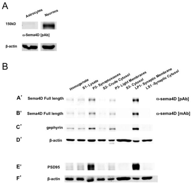
Figure 5. Sema4D is localized to the synaptic membrane.
(a) Western blot analysis of lysates from DIV16 primary cultures of astrocytes isolated from P1 rat pups or hippocampal neurons isolated from E18 rat pups.
(b) Western blot analysis of fractionated cellular lysates (see Fig S1) taken from hippocampal tissue of postnatal day 12 (P12) rat pups. (A′–D′) Western blot probed with (A′) polyclonal anti-sema4D, (B′) monoclonal anti-sema4D, (C′) anti-gephyrin, (D′) anti-β-actin. (E′–F′) Western blot analysis of same lysates as in A′–D′ probed with (E′) anti-PSD95, (F′) anti-β-actin. The same blot as in A′–D′ could not be reprobed with anti-PSD95 due to similar molecular weights of PSD95 and gephyrin.

