
Fig. 3.
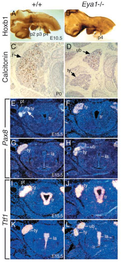
Ultimobranchial body and thyroid lobe defects in Eya1−/−mice. (A,B) The embryos of E10.5, after completion of neural crest migration, were stained with Hoxb1 antibody to label the 4th pharyngeal pouches (p4). The 4th pharyngeal pouches express Hoxb1 (brown stain) in both wild-type (A) and Eya1−/− embryos (B). No significant difference of Hoxb1 expression in the 4th pouch endoderm was observed in Eya1 mutants. (C,D) Transverse sections through newborns, stained with anti-calcitonin antibody (brown staining). (C) A wild-type thyroid with numerous calcitonin-positive cells and follicular cells (f, arrow) throughout the lobe. (D) An Eya1−/− animal with bilateral persistent ultimobranchial bodies and malformed thyroid lobes. Only a few follicles are formed in the main body of the thyroid (ty, arrow). Note that the dorsally placed vesicle which is strongly positive for calcitonin represents a persistent ultimobranchial body (ub) in Eya1-mutants, not fusing with the thyroid lobe. This ultimobranchial body also contains follicle-like structures (arrow). No isthmus was present in this animal. (E–H) Transverse sections at E15.5 showing Pax8 expression in the thyroid lobes in wild-type and Eya1−/− embryos. (E) The ultimobranchial body cell populations showing weak Pax8 expression (arrow) were visible within the thyroid lobes at this stage. Parathyroid (pt) was also visible. (F) No ultimobranchial body cell population was observed within the Eya1−/− thyroid lobes on the same level and parathyroid was also absent. However, the ultimobranchial bodies showing weak Pax8 expression were observed at the anterior end dorsal to the thyroid lobe in Eya1−/−embryos (H). No ultimobranchial bodies were found in wild-type embryos on the same level (G). (I–L) Transverse sections showing Ttf1 expression in the thyroid lobes in wild-type embryos at E15.5 (I,K) and in both the persistent ultimobranchial bodies and the thyroid lobes of Eya1−/− embryos (J,L). Similarly, the persistent ultimobranchial bodies were observed as separate structures located anterodorsally to the thyroid lobes in Eya1−/− animals (L). For C–L, dorsal is up. tr, trachea; la, larynx.