Fig. 5.
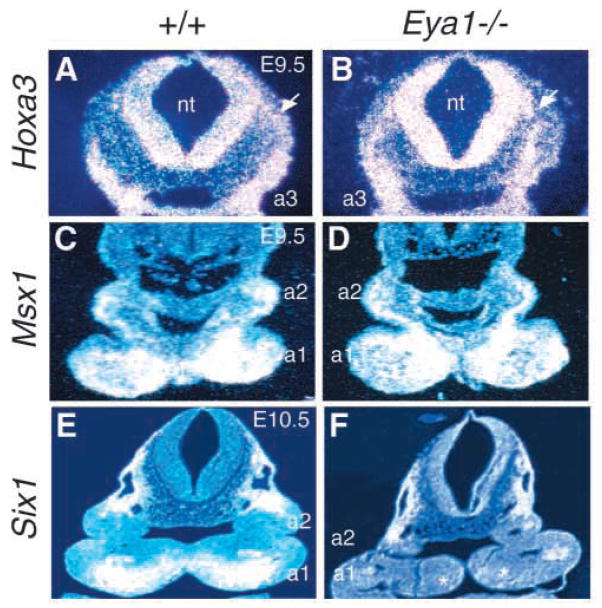
Neural crest defects in the pharyngeal arches of Eya1−/−embryos. (A,B) Transverse sections of wild-type and Eya1−/−embryos at E9.5 showing Hoxa3 expression in the hindbrain neural tube, migrating neural crest cells (arrow) and 3rd pharyngeal arches (a3). No significant change was found in Eya1 mutants. (C,D) Transverse sections showing Msx1 expression in the neural crest cells in the pharyngeal arches of wild-type and Eya1−/− embryos at E9.5. No significant difference of Msx1 expression was observed at E9.5 in Eya1−/− embryos. (E,F) Transverse sections showing Six1 expression in the distal edge of arch mesenchyme in wild-type and Eya1−/− embryos at E10.5. Six1 expression was not detectable in Eya1−/− embryos (asterisks in F). Dorsal is up.

