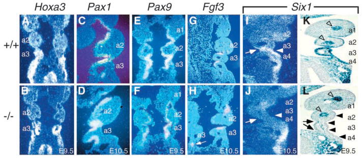
Fig. 6.

Expression of Six1 in the pharyngeal pouch endoderm and the ectoderm of pharyngeal clefts was markedly reduced in Eya1−/− embryos. (A,B) Coronal sections showing that Hoxa3 is normally expressed in the 3rd and 4th pharyngeal pouches and arches (A), and its expression was unaffected in Eya1−/− embryos at E9.5 (B). (C,D) Coronal sections showing that Pax1 is normally expressed in the pouch endoderm at E9.5–10.5 (C), and its expression is preserved in Eya1−/− embryos (D). (E,F) Coronal sections showing that Pax9 is also expressed in the pouch endoderm at E9.5–10.5 (E) and its expression is also preserved in Eya1−/− embryos (F). (G,H) Coronal sections showing that Fgf3 is normally expressed in the posterior half of the pouch endoderm at E10.5 (G) and its expression was unaffected in Eya1−/− embryos (H). (I,J) Coronal sections showing that Six1 is normally co-expressed with Eya1 in the 3rd pouch endoderm (arrow in I) and the surface ectoderm of 3rd clefts (arrowhead in I), and its expression was markedly reduced in both structures in Eya1−/− embryos (arrows and arrowheads in J). (K,L) Coronal sections showing strong Six1lacZ expression in wild type (K) in the pouch endoderm and surface ectoderm including 2nd, 3rd and 4th pharyngeal clefts; however, in Eya1−/− embryos, Six1lacZ expression was significantly reduced in the pouch endoderm (arrows) and surface ectoderm (arrowheads, L) in the 2nd, 3rd and 4th pharyngeal regions (a2–a4). In contrast, its expression surrounding the arteries of pharyngeal arches remains unaffected (open arrowheads).
