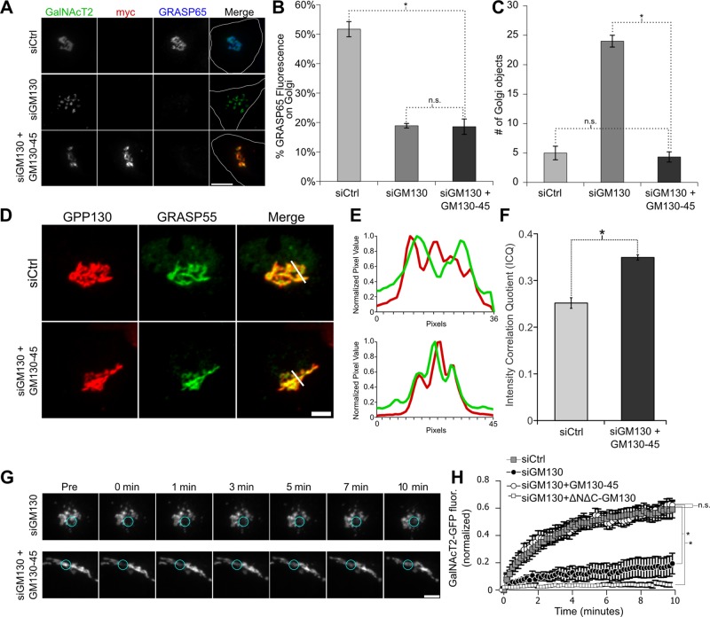
FIGURE 4:
The GM130-45 chimera rescues Golgi ribbon integrity after GM130 knockdown. (A) Immunofluorescence staining for cells stably expressing GalNAcT2-GFP that were transfected with control or GM130-specific siRNA 96 h before fixation. Where indicated, the cells were also transfected with the myc-tagged GM130-45 chimera 12 h before fixation. Scale bar, 5 μm. (B) The amount of GRASP65 fluorescence present on the Golgi was determined using GalNAcT2-GFP as a mask. The percentage of the total cellular pool that is present on the Golgi is shown for each condition (n = 10, mean ± SEM; *p < 0.05; n.s. = p > 0.05). (C) The number of Golgi objects present for each condition determined by counting discrete GalNAcT2-GFP–positive objects (n = 10, mean ± SEM; *p < 0.05; n.s., not significant). (D) Average value z-projections of GRASP55 and GPP130 immunofluorescence images in control and mycGM130g45tail-rescued–expressing cells. Expression of GM130-45 increases colocalization of GRASP55 with cis-Golgi marker GPP130, as shown by profile plots (E) and the intensity correlation quotient (F). (G) Time course of Golgi fluorescence recovery after photobleaching in siGM130 and GM130-45–rescued cells. Blue circles encompass the photobleached region. Scale bar, 5 μm. (H) Fluorescence levels in bleached region measured and plotted vs. time in siCtrl-treated cells and siGM130 cells only and siGM130 cells transfected with GM130-45 or ∆N∆C-GM130 (n = 10 cells, mean ± SEM; *p < 0.05; n.s., not significant).

