FIGURE 1:
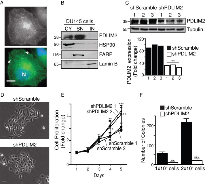
PDLIM2 suppression alters morphology, proliferation, and clonogenic growth. (A) DU145 cells were immunostained for PDLIM2 (gray/green), counterstained with Hoechst (blue; N), and photographed. Scale bar, 10 μm. Arrows indicate points of focal adhesion. (B) Subcellular fractionations of cytoplasmic (CY), soluble nuclear (SN), and insoluble nuclear (IN) fractions of DU145 cells analyzed by Western blotting for PDLIM2 expression. Purity of the fractions was confirmed using markers of each fraction: CY (HSP90), SN (PARP), and IN (lamin B). (C) Clones of DU145 cells stably expressing scrambled shRNA (shScramble) or PDLIM2 shRNA (shPDLIM2) were analyzed by Western blotting. PDLIM2 expression normalized to tubulin loading control, relative to shScramble clone2, quantified by densitometry using Odyssey software. (D) Cells were plated at low densities on collagen for 5 d, and colonies were photographed with phase microscopy. Representative micrographs of three separate experiments. Scale bar, 50 μm. (E) Cell cultures seeded at equal densities were monitored by crystal violet staining over 5 d. Data represent an average of six wells from a representative experiment. (F) Cells seeded at equal densities in soft agarose were cultured for 4 wk, when colonies were stained with crystal violet and counted. Data represent mean colonies per well from triplicate cultures (*p < 0.05, **p < 0.005, ***p < 0.0005).

