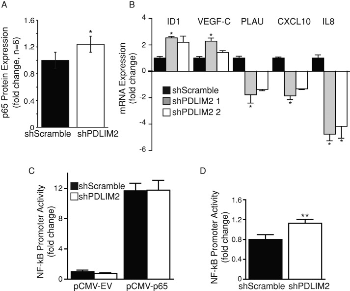
FIGURE 4:
Suppression of PDLIM2 does not alter basal NFκB activity in DU145 cells. (A) Western blotting for NFκB p65 expression in shScramble and shPDLIM2 DU145 cells. Densitometry measurements of the average fold difference in p65 expression in shPDLIM2 cells compared with shScramble controls from six independent experiments. (B) Quantitative PCR on total RNA extracted from shScramble and shPDLIM2 cells for PLAU, CXCL10, IL8, ID1, VEGF-C, and PDLIM2 mRNA expression. β2M mRNA levels were used as a control; expression levels were compared to shScramble mRNA levels and expressed as fold change. (C) shScramble or shPDLIM2 DU145 cells were transfected with a dual-luciferase plasmid containing an NFκB promoter and cotransfected with pCMV-empty vector (pCMV-EV) or pCMV-p65 construct. (D) Cells were transfected as in C and treated with 20 ng/ml TNF-α for 4 h before analysis. Luciferase activity in p65-transfected cells normalized to that of Renilla luciferase is presented as mean fold change of activity in shScramble cells expressing the empty vector ± SEM in triplicate samples and three independent experiments. *p < 0.05, **p < 0.005.

