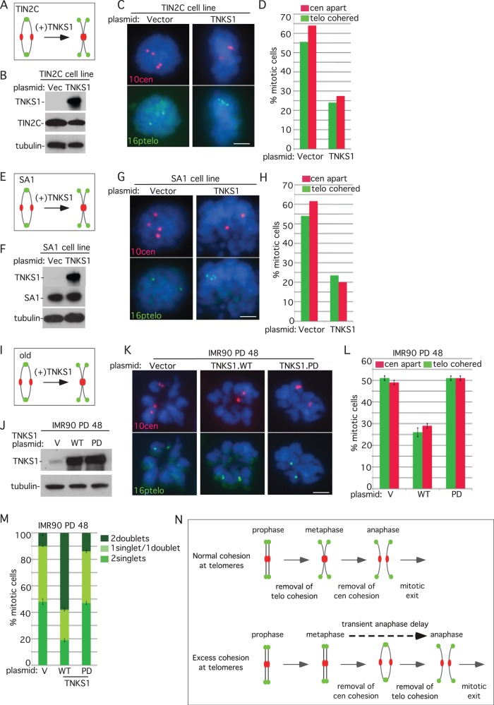
FIGURE 5:
Tankyrase 1 overexpression rescues persistent telomere cohesion, regardless of the mechanism of induction. (A–H) Tankyrase 1 overexpression rescues persistent telomere cohesion and loss of centromere cohesion in TIN2C- or SA1-overexpressing cells. (A, E) Schematic diagrams. (B–D) HTC75.V or TIN2C and (F–H) HTC75.V or SA1 cells were transfected for 24 h with a vector or TNKS1 plasmid and analyzed by (B, F) immunoblot and (C, G) centromere (red) and telomere (green) FISH after mitotic shake-off. DNA was stained with DAPI (blue). Scale bars, 5 μm. (D, H) Graphical representation of the frequency of mitotic cells with centromeres apart and telomeres cohered (D, n = 50–56 cells each; H, n = 50–60 cells each). (I–L) Wild-type but not PARP-dead tankyrase 1 rescues telomere cohesion and centromere separation in aging IMR90 cells. (I) Schematic diagram. (J–L) IMR90 cells stably expressing tankyrase 1 WT or PARP dead or a vector control were generated by lentiviral infection at an early PD (20). Cell lines were grown for multiple PDs and then, before senescence (PD 48), analyzed by (J) immunoblot and (K) centromere (red) and telomere (green) FISH after mitotic shake-off. DNA was stained with DAPI (blue). Scale bar, 5 μm. (L) Graphical representation of the frequency of mitotic cells with telomeres cohered and centromeres apart. Values are means ± SEM, derived from two independent experiments (n = 50 cells each). (M) Loss of synchrony in sister telomere separation in aging IMR90 cells can be rescued by wild-type but not PARP-dead tankyrase 1. Graphical representation of the frequency of mitotic cells with one singlet/one doublet, two singlets, or two doublets in IMR90 cells expressing vector, TNKS1.WT, or TNKS1.PARP dead at PD 48. Values are means ± SEM, derived from two independent experiments (n = 50 cells each). (N) Model showing that excess telomere cohesion leads to anaphase delay.

