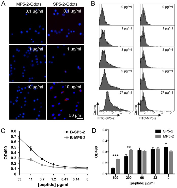
Figure 3. Verification of SP5-2 binding to a mouse (LL/2) and human (H460) lung cancer cell lines.
(A) Binding of peptide-conjugated Qdots to tumor cells in vitro is peptide-sequence specific. The ability of quantum dots labeled with SP5-2 and MP5-2 to bind to the surface of LL/2 cells was examined (Scale bar, 50 µm). Nuclear staining was performed using DAPI. (B) LL/2 cells were incubated with SP5-2 FITC-conjugated (FITC-SP5-2) or MP5-2 FITC-conjugated (FITC-MP5-2) peptides at increasing concentrations. The FITC-SP5-2 treated LL/2 cells showed increasing flurescence intensity at increasing peptide concentrations, but no such correlation was observed in FITC-MP5-2 treated cells. (C) H460 cells were seeded in 96-well ELISA plates. Biotinylated SP5-2 (B-SP5-2) and biotinylated MP5-2 (B-MP5-2) were used to test the peptide binding assay. (D) Peptide competitive inhibition assay with the unlabeled SP5-2 peptide. The black bars showed the binding inhibition of the biotinylated peptide (B-SP5-2, 10 µg/ml) by the unlabeled SP5-2 peptide (from 22 to 600 µg/ml). The gray bars showed the binding inhibition of B-SP5-2 by the MP5-2 peptide. The statistic value was compared to control peptide. **, p value <0.01; ***, p value <0.005.

